
Recently scientists and researchers at Johns Hopkins University developed an assessment technology that is much better at predicting arrhythmia risk than the blood pumping test. The team performed pre-implant MRIs from patients who were determined to need defibrillator implants. They used the MRIs to create personalized, virtual 3-D hearts. They then used the 3-D models to simulate the electrical processes in the heart to predict the likelihood that the virtual heart would develop arrhythmia. If the virtual heart-based predictions were accurate they could use the non-invasive MRI to rule out the invasive implant. The technology is called VARP, for virtual heart arrhythmia risk predictor.
When electrical waves in the heart run amok in a condition called arrhythmia, sudden death can occur. To save the life of a patient at risk, doctors currently implant a small defibrillator to sense the onset of arrhythmia and jolt the heart back to a normal rhythm. But a thorny question remains: How should doctors decide which patients truly need an invasive, costly electrical implant that is not without health risks of its own?
Trayanova, a pioneer in developing personalized imaging-based computer models of the heart, supervised the research and was senior author of the journal article. She holds faculty appointments within Johns Hopkins’ Whiting School of Engineering and itsSchool of Medicine, and she is a core faculty member of the university’s Institute for Computational Medicine. For this study, she joined forces with cardiologist and co-author Katherine C. Wu, associate professor in the Johns Hopkins School of Medicine, whose research has focused on magnetic resonance imaging approaches to improving cardiovascular risk prediction.
For this landmark study, Trayanova’s team formed its predictions by using the distinctive magnetic resonance imaging (MRI) records of patients who had survived a heart attack but were left with damaged cardiac tissue that predisposes the heart to deadly arrhythmias. The research was a blinded study, meaning that the team members did not know until afterward how closely their forecasts matched what happened to the patients in real life. This study involved data from 41 patients who had survived a heart attack and had an ejection fraction—a measure of how much blood is being pumped out of the heart—of less than 35 percent.
To protect against future arrhythmias, physicians typically recommend implantable defibrillators for all patients in this range, and all 41 patients in the study received the implants because of their ejection fraction scores. But research has concluded that this score is a flawed measure for predicting which patients face a high risk of sudden cardiac death.
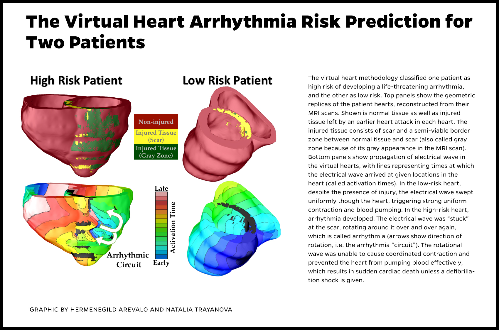
The Johns Hopkins team invented an alternative to these scores by using pre-implant MRI scans of the recipients’ hearts to build patient-specific digital replicas of the organs. Using computer-modeling techniques developed in Trayanova’s lab, the geometrical replica of each patient’s heart was brought to life by incorporating representations of the electrical processes in the cardiac cells and the communication among cells. In some cases, the virtual heart developed an arrhythmia, and in others it did not. The result, a non-invasive way to gauge the risk of sudden cardiac death due to arrhythmia, was dubbed VARP, short for virtual-heart arrhythmia risk predictor. The method allowed the researchers to factor in the geometry of the patient’s heart, the way electrical waves move through it and the impact of scar tissue left by the earlier heart attack.
Eventually, the VARP results were compared to the defibrillator recipients’ post-implantation records to determine how well the technology predicted which patients would experience the life-threatening arrhythmias that were detected and halted by their implanted devices. Patients who tested positive for arrhythmia risk by VARP were four times more likely to develop arrhythmia than those who tested negative. Furthermore, VARP predicted arrhythmia occurrence in patients four-to-five times better than the ejection fraction and other existing clinical risk predictors, both non-invasive and invasive.