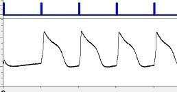
Photoswitches are a family of small-molecules designed to photo-modulate the activity of specific signaling proteins: One color turns the protein ‘on’; a different color turns it ‘off’. Photoswitches were conceived at the Nanomedicine Development Center for the Optical Control of Biological Function at UC Berkeley
Light-sensitive proteins are changing the way scientists study how new drug candidates affect critical properties of heart and nerve cells. The proteins, discovered by Max Planck researchers, are incorporated into model cells and, with an instrument developed by Photoswitch Biosciences, used to control the function of other ion channels. Monitoring tiny electrical changes in the cells allows researchers to screen chemical libraries for new drug candidates or to evaluate the safety of new drugs for use in humans.

Ion channels are the third largest pharmaceutical target class. The discovery and development of ion channel drugs has been hampered by a lack of cost-effective, physiologically relevant, high-throughput assays. Using light to both control membrane potential and readout ion channel activity enables an entirely new ion-channel assay paradigm with dramatically highter throughput and lower cost than existing technologies. Photoswitch Biosciences is developing a complete assay system consisting of cell lines, photoswitches, voltage-sensitive dyes and a reader.
Retinitis pigmentosa (RP) is an inherited, blinding disease, affecting approximately 100,000 people in the US. There are no approved treatments.
Persons with RP lose the light-sensitive rods and cones at the back of the eye. Our goal is to render the still healthy neural cells of the retina light sensitive. Those photoswitched retinal cells would assume the role of the degenerated photoreceptors. Photoswitches have been shown to restore light sensitivity to the retina in mouse models of RP.

In 2002, Max Planck researchers first isolated Channelrhodopsins, from the green algae Chlamydomonas reiinhardii, and successfully used both in vitro andin vivo to control and study a variety of critical bioelectrical systems. Now the US company Photoswitch Biosciences is developing a complete assay system specifically designed for drug screening and safety pharmacology studies. This enables researchers to make use of the benefits of channel rhodopsin-based optical control. The system analyses the function of heart and nerve significantly faster and more cost-effectively than previous methods.
The new instrumentation platform was developed partly on the basis of a non-exclusive license agreement with Max Planck Innovation, the technology transfer organization of the Max Planck Society, for the use of biological photoreceptors for the direct control of light activated ion channels.
http://www.photoswitchbio.com/index.html
